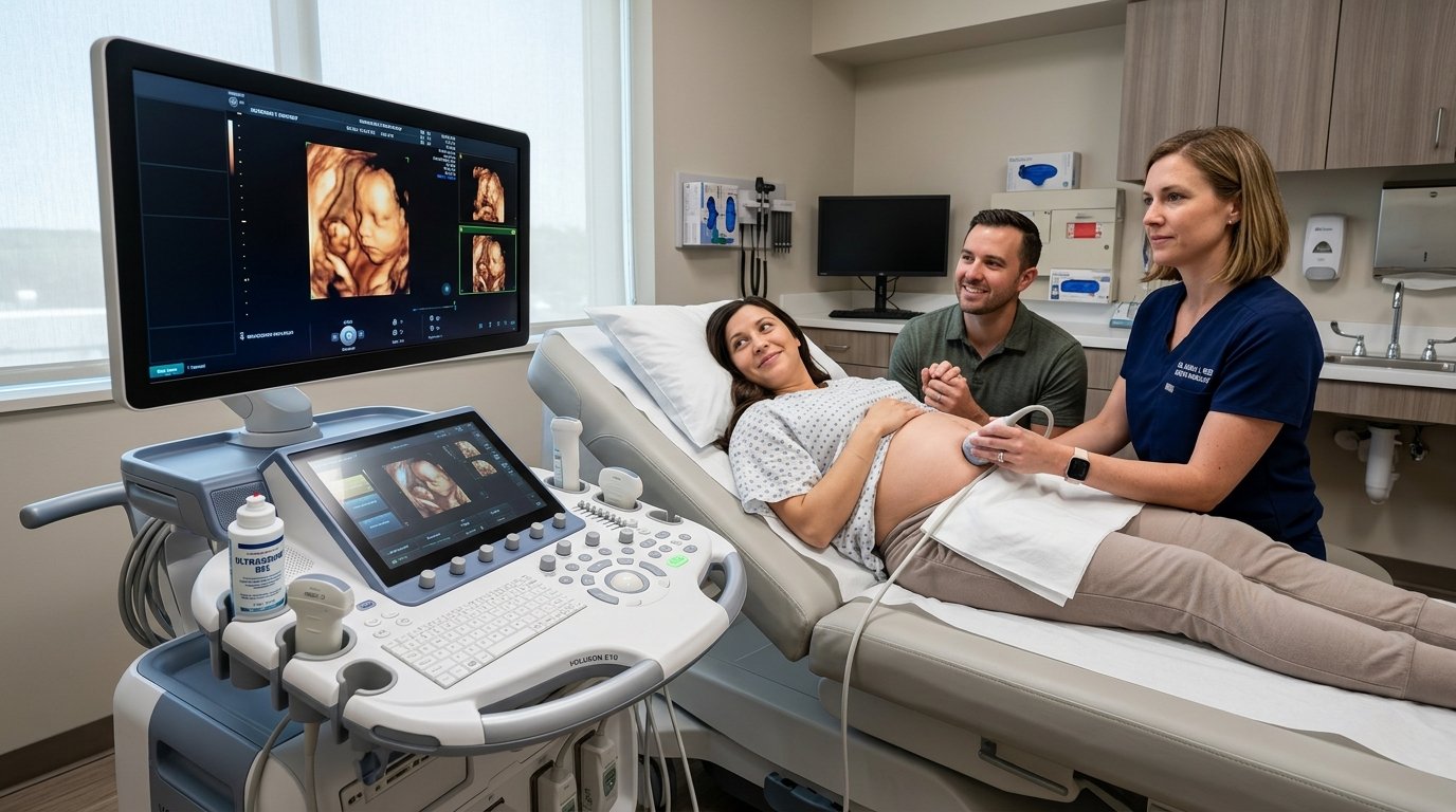
The Evolution of Obstetric Ultrasound: Beyond 2D Imaging
Four-dimensional (4D) obstetric ultrasound represents a major leap in modern fetal medicine. While conventional 2D ultrasound provides cross-sectional planar images critical for anatomical biometry, 4D ultrasound captures high-resolution volume rendering with real-time motion, providing parents and obstetricians with a vivid dynamic window into fetal development.
Optimal Timing for a 4D Ultrasound Scan
Although 4D imaging can be performed across various stages of gestation, the ideal diagnostic window is between weeks 26 and 32 of pregnancy:
- Weeks 26 to 28: Fetal facial features are well-delineated with adequate subcutaneous fat, and there is ample amniotic fluid allowing unobstructed views.
- Weeks 29 to 32: Fetal movements, facial grimaces, sucking, and smiling can be observed in vivid detail.
- Beyond Week 34: As the fetus grows and occupies the majority of the uterine cavity, descent into the pelvis or reduced relative fluid volume may make full face visualization more challenging.
Clinical and Emotional Benefits
- Congenital Anomaly Screening: Facilitates detailed surface evaluation of cleft lip, palate defects, cranial sutures, and limb micro-anomalies.
- Placental and Amniotic Assessment: Evaluates placental architecture, cord insertion, and fluid index alongside Color Doppler hemodynamic analysis.
- Maternal-Fetal Bonding: Seeing real-time fetal movements and facial expressions fosters deep emotional connection and reduces maternal pregnancy anxiety.
Patient Tip: Drink plenty of water in the days leading up to your ultrasound appointment to optimize amniotic fluid clarity, and enjoy a light snack 30 minutes before your scan to encourage gentle fetal movement.